Stratasys and Siemens Healthineers recently presented the results of a joint research effort that demonstrates the unprecedented accuracy of the solutions offered by Stratasys to enable 3D-printed medical imaging phantoms to replicate human anatomy.
This collaborative effort uses Stratasys’ RadioMatrix materials and Digital Anatomy technology with Siemens Healthineers’ advanced algorithms to improve the quality of complex anatomy medical imaging phantoms, allowing surgeons, researchers, and educators to replace simplistic anatomical phantom models for pre-surgery planning and education.
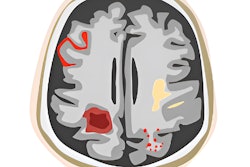
This new research demonstrates how anatomically accurate, patient-specific 3D-printed phantoms are scalable, cost-effective and efficient in developing new CT scan algorithms and improving diagnostic precision. By using 3D-printed anatomic models for radiology, it is possible to offer a patient-specific, anatomical model that accurately replicates anatomy and pathologies.
The results of the study were shared during this winter’s Radiological Society of North America (RSNA) annual meeting.
The joint efforts will drive innovation in medical imaging through multiple ways:
- Integrating Stratasys’ advanced Digital Anatomy technology and materials with Siemens Healthineers’ imaging creates patient-specific phantoms with realistic radiopacity and anatomical precision. These models replicate clinical imaging outcomes more closely than existing traditional phantoms, addressing the radiology field’s long-standing need for consistency and reliability while preserving the anatomy and details of potential pathologies and/or variations of anatomy.
- With the ability to produce repeatable data sets on the same anatomy, these phantoms eliminate ethical, and variability challenges associated with human scans and/or the usage of cadavers.
- Advanced post-processing including validation data for AI-based software solutions. The ultra-realistic phantoms accelerate the development of imaging algorithms, driving materials innovation, and enabling the exploration of new clinical and academic applications.
By utilization of these kind of 3D-printed phantoms, hospitals and imaging centers can enhance the calibration and performance of CT scanners, ensuring more accurate diagnostics and better patient outcomes. These phantoms are also opening new doors for education, training, and research in radiology, ultimately improving patient outcomes and costs.
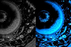
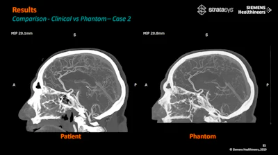
The RSNA presentation also highlighted the significant deviations between real and printed models, with variances as low as single Hounsfield units (HU) in critical areas like grey matter and veins. This extraordinary level of accuracy redefines the standards for CT imaging research.