An estimated five to ten percent of blindness worldwide is caused by the rare inflammatory eye disease uveitis. Posterior uveitis in particular is often associated with severe disease progression and the need for immunosuppressive therapy.
In posterior uveitis, inflammation occurs in the retina and in the underlying choroid that supplies it with nutrients.
Researchers at the Ophthalmology Department at the University of Bonn have tested color-coded fundus autofluorescence as a supportive novel diagnostic method.
Fluorescence of the retina can be used to infer the uveitis subtype. This is an essential prerequisite for accurate diagnosis and treatment of the disease. The results have now been published in Scientific Reports.
Blurred vision, floaters and unusual light perception - those affected by the rare disease posterior uveitis feel no pain. "But the consequences can be severe: About five to ten percent of blindness worldwide is caused by uveitis. Uveitis is a rare disease, but posterior uveitis in particular potentially has a poor prognosis and often requires immunosuppressive therapy," explains Dr. Maximilian Wintergerst of the Ophthalmology Department at the University of Bonn. There are different forms of the disease. In posterior uveitis, the retina or choroid in the eye becomes inflamed. While the retina converts the incident light into nerve impulses, the choroid supplies the outer layers of the retina with nutrients.
Different therapeutic management
"It's not easy to distinguish between the numerous subtypes of uveitis," says Wintergerst. However, since the different subtypes often require a different therapeutic approach, a reliable diagnosis is all the more important. This is why researchers from the Ophthalmology Department at the University of Bonn, together with colleagues from the Medical Biometry and Rheumatology Departments at the University Hospital Bonn and the University Hospital of Ophthalmology in Bern (Switzerland), investigated a new imaging technique that can assist in the diagnosis of posterior uveitis.
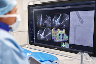
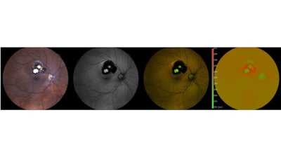
The team evaluated color-coded fundus autofluorescence (Spectrally Resolved Autofluorescence Imaging). The CenterVue (iCare) company from Padua (Italy) provided the newly developed device to the researchers for the examinations. This process involves illuminating the retina with bluish light. The retina absorbs the light and emits it again at a different wavelength. The device measures this fluorescence and splits the signals into a green and a red component.
"The green-to-red ratio of the light emitted from each inflammatory focus depends, among other factors, on the exact posterior uveitis subtype involved," Wintergerst explains. The researchers examined the eyes of 45 study participants. In all of them, the exact subtype of uveitis was diagnosed beforehand. This included ophthalmologic examination findings, laboratory investigations, serologic and radiologic findings, and in some cases genetic and interdisciplinary clinical examinations.
More reliable diagnoses with color-coded fundus autofluorescence
The researchers evaluated the green-red ratio in the fluorescence of the fundus of the eye for about 800 inflammatory foci in the eyes of the patients. "Our results indicate, that this ratio can be very characteristic and helpful as a marker for differentiating the various posterior uveitis subtypes," states Prof. Dr. Dr. Robert Finger, co-author of the study and head of the uveitis clinic at the Ophthalmology Department at the University Hospital Bonn. "It could allow us to make more confident diagnoses in the future." This is a big step for the Ophthalmology Department at the University of Bonn, especially since Finger coordinates the Germany-wide uveitis registry "TOFU" (Treatment exit options for non-infectious uveitis, www.tofu-uveitis-register.de) together with colleagues from Münster. The aim is to document disease progression on a long-term basis and to develop recommendations for treatment guidelines.
"In the current study, we present the precise technical background of color-coded fundus autofluorescence in ophthalmology in collaboration with our international partners," says head of department Prof. Dr. Frank Holz. "This technology may also allow better monitoring of posterior uveitis in the future, in addition to more reliable diagnoses."
The study was funded by the BONFOR-GEROK program of the Faculty of Medicine of the University of Bonn.