A seizure is a sudden, overwhelming wave of electrical energy passing through the brain in a matter of seconds — so fast, in fact, that it’s been nearly impossible to fully capture where these electrical bursts originate, how they move and where they stop.
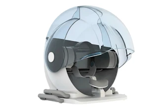
That’s why University of Georgia researchers developed a new high-resolution light-sheet imaging system that is fast enough to document seizures in 3D in real-time.
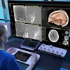
Using zebrafish larvae, the go-to animal model for neuroscience research, the researchers captured images of a seizure making its way through the brain.
The images show that the seizure began toward the back of the brain and moved forward toward the part of the midbrain that processes visual information known as the optic tecta. This region controls eye movement and manages responses to what the animal is seeing. The electrical activity gradually subsides over several seconds.
The study is among the first to provide a high-resolution 3D video of a seizure event from start to finish.
 UGA
UGA
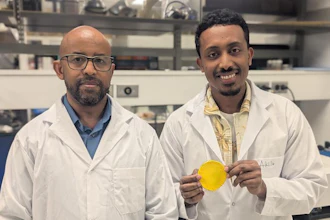
“The brain is obviously three dimensional, so when you have 2D imaging, not everything is going to be visible on that single 2D plane,” said Peter Kner, corresponding author of the study and a professor in UGA’s College of Engineering. “Seeing where something is going or where something happens, if you’re looking at a 2D plane, you start to wonder, ‘Did I actually capture the whole thing?’”
Examining those images in 3D could offer new insights into how seizures form and how they propagate. Seizure propagation is how seizure activity starts in one part of the brain and moves through other parts of the brain. This, in turn, helps researchers understand how the brain operates.
A better understanding of how the brain operates could inform new treatments of brain diseases and disorders, Kner said.
Light sheet microscopy uses a thin sheet of light to illuminate a single slice of a sample at one time. It works well on living organisms because it provides clear images at high-speed with low background, enabling fast tracking of complex processes like brain activity.
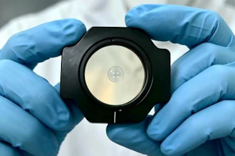
The new microscope also relies on adaptive optics, a technology originally developed for use in astronomy.
“When you look at the stars in the night sky, they sort of twinkle because the atmosphere is making the image wobble around,” Kner explained. “It looks nice, but it’s not great for astronomers because they don’t get a good, sharp image. Adaptive optics technology corrects that.”
A similar problem is present in brain imaging caused by the tissues the light travels through. As the light travels through the tissue, its path gets bent, and the images get blurred. Using adaptive optics enables researchers to get a sharper image.
“You always want the sharpest image you can get,” Kner said. “The whole field of imaging is really exciting right now. Microscopes have been around since roughly 1650, so you think what could possibly be new?
“But there are a lot of places left for the field to go.”
Published by Biomedical Optics Express, from the Optica Publishing Group, the study was funded by a grant from the National Institutes of Health. It was co-authored by Bingxi Liu and Yang Liu, who both graduated from UGA with doctorates in electrical and computer engineering; Carly Duffy, who received a doctorate from UGA’s department of cellular biology; and James Lauderdale, department head and professor of cellular biology.