Amsterdam, the Netherlands – Royal Philips, a health technology company, has announced the introduction of its VeriSight Pro 3D Intracardiac Echocardiography (ICE) catheter in Europe. Building on its success in the United States, VeriSight Pro brings real-time 3D imaging directly inside the heart, helping physicians perform procedures with greater clarity — without the need for general anesthesia.
Designed for procedures such as transcatheter valve repair and left atrial appendage closure, VeriSight Pro offers high-resolution 2D and 3D visualization directly within the heart chambers. This enables clinical decision-making in structural heart and electrophysiology interventions, while removing the need for general anesthesia and associated risks.
Prof. Dr. Jörg Hausleiter, Ludwig-Maximilians-Universität (LMU) Munich, Germany, said, “With VeriSight Pro 3D ICE, we now have the ability to see detailed cardiac anatomy from inside the heart in real time. This helps streamline our workflows and makes complex procedures more accessible to patients who may not tolerate more invasive imaging approaches.”
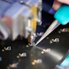
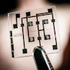
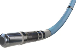
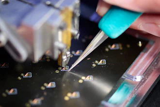
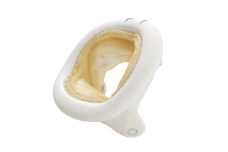
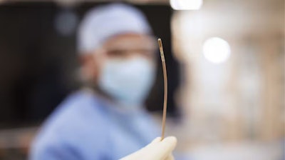
VeriSight Pro is a miniaturized ultrasound probe, approximately 3 mm in diameter, embedded at the tip of a thin, steerable catheter. The tiny device can be navigated through the vascular system and into the heart chambers, where it delivers high-quality 2D and 3D images in real time. Imaging the heart from within, with control over the scan angle, opens new possibilities for guiding structural heart interventions.
Physicians can assess anatomy, guide device placement, and confirm procedural results — all from a single access point, and without the need for more invasive imaging techniques. As the first ICE catheter to miniaturize the same 3D imaging technology used in TEE, VeriSight Pro helps address key barriers in delivering efficient, scalable care — from patient tolerance to resource availability in interventional suites.
The catheter’s unique features — including xPlane and iRotate technologies — allow physicians to visualize two imaging planes simultaneously and digitally adjust views without physically repositioning the catheter tip. These capabilities enable assessment and device deployment with fewer imaging steps.