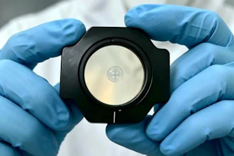
Stratasys announced full commercial availability of its RadioMatrix radiopaque 3D printing material in the United States.
This follows initial limited deployments and marks the first time healthcare providers, device manufacturers, and research institutions across the U.S. can broadly access and use the material for medical imaging and training applications.
The company said RadioMatrix is the first 3D printing material that enables control of radiopacity - allowing users to produce patient-specific models with repeatable, consistent, and fully tunable visibility on X-ray based imaging.
Its improved fidelity for computed tomography (CT) phantoms was later underscored by research conducted with Siemens Healthineers, which validated RadioMatrix’s capabilities and accuracy for accelerating innovation in device testing, calibration, and education.
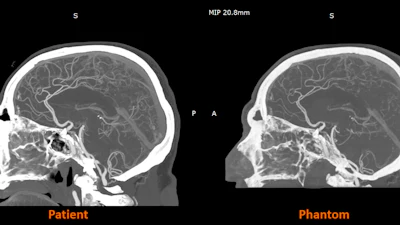
UK-based work with partners such as CPI and Beaumont Hospital is already demonstrating the impact of radiopaque 3D-printed models in practice, with radio-realistic cerebral angiography phantoms being used to improve the fidelity of imaging-based training and create more controlled, repeatable environments for research.
Early research from the Stratasys–Siemens Healthineers collaboration shows that 3D printed RadioMatrix phantoms can closely replicate real human tissue in CT imaging, with deviations reported as low as single Hounsfield units (HU) in critical areas such as grey matter and veins. By combining Stratasys’ Digital Anatomy 3D printing technology and radiopaque materials with advanced imaging algorithms, the partners are demonstrating anatomically realistic, radio-accurate phantoms that preserve fine anatomical details and pathological variations while offering a more consistent, ethical alternative to cadavers. These models are expected to improve how radiologists validate and optimize CT protocols and accelerate the development of new imaging algorithms for more precise diagnosis and treatment planning.