UNIVERSITY PARK, Pa. — Sepsis, a life-threatening condition that stems from the body trying to fight off an infection, plagues over 1.5 million patients a year in the U.S. alone, with one in three deaths recorded in the hospital attributed in part to sepsis. Despite the severity of the condition, which can kill in as little as 12 hours, it takes between two and seven days for most traditional approaches to definitively identify sepsis-causing bacteria in blood infections.
A team led by an engineer at Penn State has developed a way to condense the days-long diagnosis timeline to just hours. The new approach rapidly grows the bacteria present in collected blood samples, intermittently analyzing the samples with advanced techniques that help scientists identify the specific pathogens causing infection. The researchers reported in a paper published today (Aug. 26) in Science Advances that their approach facilitated faster diagnosis and could help clinical decision-making that avoids worsening antibiotic resistance in the bacterial strains causing infections.
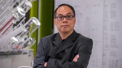
“For a physician, ‘what actually caused this infection and how should it be treated?’ are the most important questions when it comes to the timely management of a bloodstream infection,” said corresponding author Pak Kin Wong, professor of biomedical engineering and of mechanical engineering. “We are developing a comprehensive diagnostic platform that rapidly tells physicians both the specific bacteria causing a bloodstream infection, as well as the ideal antibiotic to treat the infection, before sepsis ever sets in.”
 The figure showcases the “broth” used to monitor red blood cell depletion in whole blood spiked with one colony-forming-unit of E. coli bacteria, each incubated at different orbital shaking speeds — left to right: 0 RPM, 65 RPM, 120 RPM and 200 RPM — after four hours of incubation. This culturing raises a bacteria-rich, plasma-like layer of bacteria to the top of the vials, while clusters of stuck blood cells known as a Rouleaux formation sink to the bottom.Pak Kin Wong
With a bloodstream infection, it is critical to find and neutralize the cause as quickly as possible, before it triggers a septic response from the body, Wong explained. Physicians must not only detect the presence of bacteria — they must also identify the specific pathogens, as well as the best antibiotic for treatment. The dire stakes of a false positive or negative complicate this further, as every hour counts when treating a bloodstream infection, Wong said.
“This is not like a COVID test, where we are checking to see a specific virus is present in a patient’s system,” Wong explained. “Many different bacteria can cause sepsis, and they may respond differently to treatment. Therefore, analysis must be thorough to ensure the best treatment is prescribed.”
Bloodstream infections are responsible for about 40% of all sepsis cases that lead to hospitalization. The complex biological makeup of the blood and the low pathogen loads needed to trigger sepsis make pinning the cause of a bloodstream infection time-consuming. To identify the bacteria causing a bloodstream infection, current best practices require bacterial culturing: Blood samples are enriched over a few days so that present bacteria grow to measurable levels. Then, technicians further analyze the samples to identify the specific bacteria, a process that adds another day or two to diagnosis.
Streamlining bloodstream infection diagnosis is not a novel idea, with several commercial products offering culture-free blood testing already on the market. However, to reduce diagnostic time, these products provide less comprehensive and less sensitive readings, Wong said.
To accelerate diagnosis without sacrificing accuracy, the team had to rethink culturing. Traditionally, bacterial growth in a cultured blood sample is measured through the carbon dioxide released by the bacteria. When this change in carbon dioxide levels confirms the presence of pathogens, bacteria are separated from the blood sample and analyzed. The team’s new approach, called STREAM, fast-tracks this culturing by facilitating rapid bacterial growth, while isolating and analyzing the pathogens inside simultaneously — blood samples are mixed in a specialized “broth” that separates whole blood cells from the individual bacteria found in the sample during culturing.
Molecular analysis, a process known formally as barcoding, allows the team to detect tiny fragments of genetic information from isolated bacteria. From these smaller samples collected intermittently during culturing, the researchers can name the specific bacterial species present.
“Instead of sampling at a particular endpoint after culture, we collect samples at multiple time points throughout the culture process,” Wong said. “This approach maintains robust bacterial detection while minimizing the time to results.”
These smaller samples are then subjected to a series of new, single-cell-based techniques that allow researchers to analyze a bacterium with microscopic imaging. These images are then analyzed by computer algorithms the team developed to eliminate visual clutter from the images, helping physicians determine the specific bacteria causing infection. These analyses also suggest which antibiotics the strain is susceptible to and any existing antibiotic resistance the strain may have.
The team tested their approach with about 100 positive bloodstream infection samples donated by patients and stored at Penn State Hershey Medical Center’s clinical microbiology laboratory. The researchers found that combining these techniques offered comprehensive diagnosis in as little as seven hours, enabling confident identification of the infection causing pathogen from whole blood.
“Using single-cell analysis technologies developed by our team, we can accurately identify pathogens in blood even when they are present at very low concentrations,” Wong said. “Identifying the pathogen alone is not enough; we must also determine which antibiotics are effective against it. This led us to integrate antibiotic susceptibility testing into the same process, providing physicians with the information needed to select the most appropriate treatment.”
Wong said it is important to note that just over 4% of these samples were classified as "very major errors," meaning a bacterium was inaccurately identified as resistant or susceptible to an antibiotic, which could cause the prescribed treatment to be ineffective. The team acknowledged that although some large hurdles need to be addressed before clinical implementation — including this relatively high major error rate and difficulties that could arise when testing blood from patients who had already received antibiotics — the framework offers a promising foundation for detecting and treating a leading cause of death around the world faster than ever before.
“We are integrating artificial intelligence and lab automation to make the entire process even more efficient and accurate,” Wong said. “This framework is scalable, so the list of pathogens we can detect could feasibly be expanded. We believe we could adapt this approach to identify infections originating from sources other than bacteria, like fungal infections. We hope to work closely with physicians at Penn State College of Medicine to move towards more clinical studies and, eventually, clinical adoption.”
Other co-authors affiliated with Penn State include April M. Bobenchik, associate professor of pathology and laboratory medicine; Siew Mei Chin, a biomedical engineering doctoral candidate at the time of work who has since graduated and is now a postdoctoral researcher at Stanford University’s School of Medicine; Evgenii Kovtunov, a postdoctoral researcher at the time of the work who is now a medical and public health microbiology fellow at the University of Rochester; and Emma Epiphaniou, an undergraduate researcher at the time of the work who earned her bachelor’s degree in biomedical engineering from Penn State.
Additional co-authors include Samuel Yang, professor of emergency medicine; Joseph C. Liao, professor of urology; and Kathleen E. Mach, a senior research scientist, all with Stanford University’s School of Medicine.
This work was supported by the One Health Microbiome Center Interdisciplinary Innovation Fellowship and the Leighton Riess Graduate Fellowship, as well as the National Institutes of Health’s National Institute of Allergy and Infectious Diseases under award number R01AI153133. The content is solely the responsibility of the authors and does not necessarily represent the official views of the funders.